GRADE 8 SCIENCE · FOLLOW THE LIGHT

The human eye

Follow light through the eye and distinguish optical focusing from the nerve signals that make vision possible.

Class notes

Open PDF →

Select a page to enlarge it. The PDF contains the complete set.

Follow the light pathway

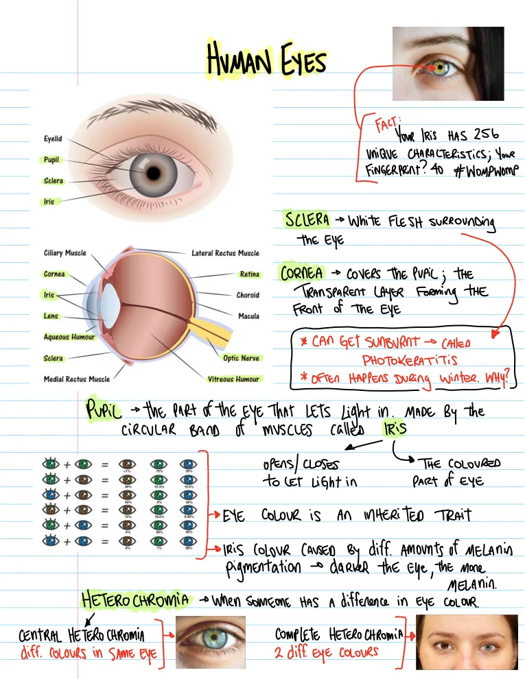
Light crosses the cornea, passes through the pupil and lens, and travels through transparent internal media toward the retina. The cornea supplies much of the focusing power. The lens adjusts focus so a sharp image can fall on the retina. The sclera is the tough outer coat.

Separate opening from controller

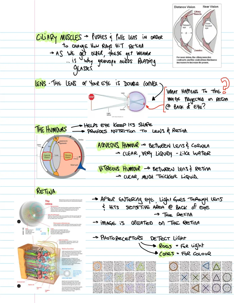
The pupil is an opening. The iris is the coloured structure with muscles that change pupil size, regulating incoming light. The aqueous humour fills the front chamber; the vitreous is a transparent gel behind the lens. These structures have different roles and should not be treated as interchangeable.

Focus changes with distance

Ciliary muscle activity and tension in supporting fibres change the lens shape. Near viewing needs increased converging power. Age-related loss of near focusing is strongly associated with lens stiffening, rather than simply “weak eye muscles.” A ray diagram represents optical focusing, not the entire visual process.

Detection is not a tiny picture sent along a nerve

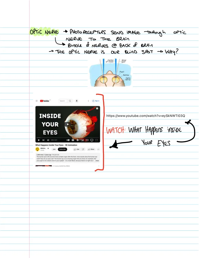
Photoreceptors respond to light; retinal circuits process information and ganglion-cell fibres carry signals through the optic nerve. The brain interprets these signals. Rods support dim-light vision; cones support colour and fine detail under brighter conditions. The optic disc lacks photoreceptors, producing a blind spot.

Words worth knowing
Accommodation
Adjustment of lens shape to change focusing power.
Retina
Light-sensitive tissue at the back of the eye.
Photoreceptor
A light-detecting cell; rods and cones are photoreceptor types.
Optic nerve
Carries visual signals from the retina toward the brain.
Learning goal

Connect each major eye structure to its function and explain accommodation and the blind spot.

WATCH THE PHENOMENON

Follow light through the eye

National Eye Institute, NIH · Anatomy animation

From an optical image to signals

Trace cornea → pupil → lens → retina, then identify where the explanation becomes about neural signals.

Open on YouTube →
PREDICT · CHANGE · EXPLAIN

Keep the image on the retina

Bring the object closer, adjust accommodation and compare a relaxed, short-sighted or long-sighted model eye.

Reduced, thin-lens eye: retina 17 mm behind the combined focusing surface; the two refractive-error models differ by ±4 D. Dioptres measure optical power. The sharpness preview is schematic.

FOLLOW THE REASONING

Work it through

Explain why the pupil gets smaller in bright conditions.

  1. The pupil is the opening through which light enters.
  2. Iris muscles change the opening size.
  3. A smaller pupil reduces the amount entering; it does not turn off the retina.

Explain a blind-spot observation.

  1. Keep one eye closed and fix attention on one mark.
  2. At an appropriate distance another mark can fall on the optic-disc region.
  3. It disappears because that region has no photoreceptors.
  4. Maintain fixation: moving the eye changes where the mark lands.
TRY IT

Try it yourself

Which controls pupil size? State iris or retina and explain your reasoning.

Give me a hint

Choose the structure around the opening.

Compare with a worked response

The iris changes pupil size.

Which tissue detects light at the back of the eye? State retina or sclera and explain your reasoning.

Give me a hint

Look for photoreceptors.

Compare with a worked response

The retina contains light-detecting cells.

TAKE IT FURTHER

Make the connection

Why is this not evidence that light stopped reaching the eye?

Compare your reasoning

The dot’s image can fall on the optic disc, where the optic nerve exits and there are no photoreceptors. The light path still exists; detection differs at that retinal location.

Common question: “The pupil is a black muscle.”

The pupil is an opening. The iris around it contains the muscles that change its size.

CHECK YOUR UNDERSTANDING

Put the ideas to work

Commit to an answer, then use the feedback to check your reasoning.

1. Accommodation changes…

2. Why is there a blind spot?

3. What travels along the optic nerve?

What are you curious about?

Search lessons, class notes and interesting questions.